Novel assembly of a head-trunk interface in the sister group of jawed vertebrates
– Were the vertebrate jaws a leader or a follower in the transformation toward 'gnathostomes'? –
Tetsuto Miyashita, Philippe Janvier, Kristen Tietjen, Feisa Berenguer, Sebastian Schöder, Federica Marone, Pierre Gueriau & Michael I. Coates
Nature DOI: 10.1038/s41586-025-09329-9
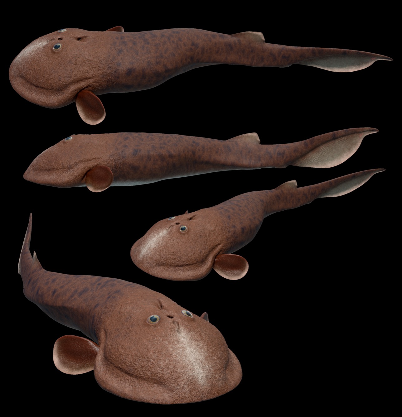
Life reconstruction of Norselaspis by Kristen Tietjen.
Also see my guest post for Springer Nature Research Community:
Paradox Lost
Out of the pandemonium of early vertebrate research emerges Norselaspis, offering an exquisite view of the dawn of jawed fishes
A Japanese expression for being fired at job literally translates to "being decapitated". Though hair-raising, the origin for this expression has little to do with the death penalty. Rather, it is rooted in a traditional puppet theatre . . . [CONTINUE READING HERE]
Executive summary
As a sister group of jawed vertebrates, osteostracans inform pre-'gnathostome' condition. Classical reconstructions of the internal anatomy – particularly that of Norselaspis – depict them as primitive, perhaps lamprey-like. This predicts that many derived traits of jawed vertebrates evolved in tandem with jaws.
Our new reconstruction of Norselaspis now show that such derived traits – many aspects of elaborate sensory systems, increased cardiac output, and greater locomotory control – were already in place in the common ancestors between osteostracans and jawed vertebrates. This means that the vertebrate jaw was a follower to and not a facilitator of these transformations.
Morphological transformations across the origin of jawed vertebrates concern more than just the feeding apparatus. We highlight the interface between the head and trunk as the centre of these character changes.
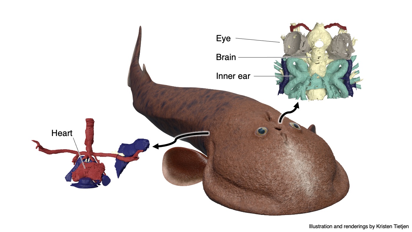
Visual summary
Highlights from the Paper
- We synchrotron-scanned the osteostracan Norselaspis to reconstruct its internal anatomy.
- Norselaspis has a much taller vertical profile than originally reconstructed in 1981.
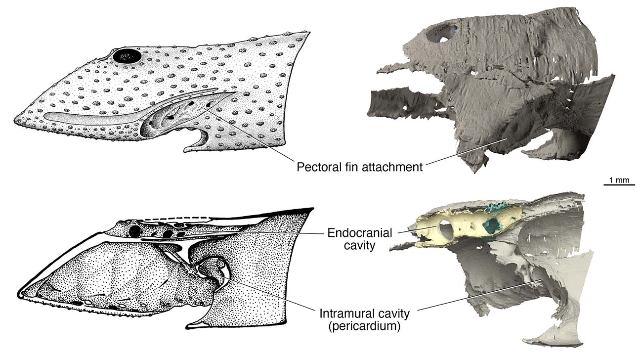
Left: The original reconstruction of Norselaspis by Janvier (1981) in lateral view (upper) and sagittal section (lower). Drawing by Philippe Janvier.
Right: Our new reconstruction of Norselaspis in lateral view (upper) and sagittal section (lower). Rendering by Kristen Tietjen.
- The inner ear is proportionally large, occupying just about half the length of the entire endocast. Pars inferior accounts for half the labyrinthian height, filled with otoconia.
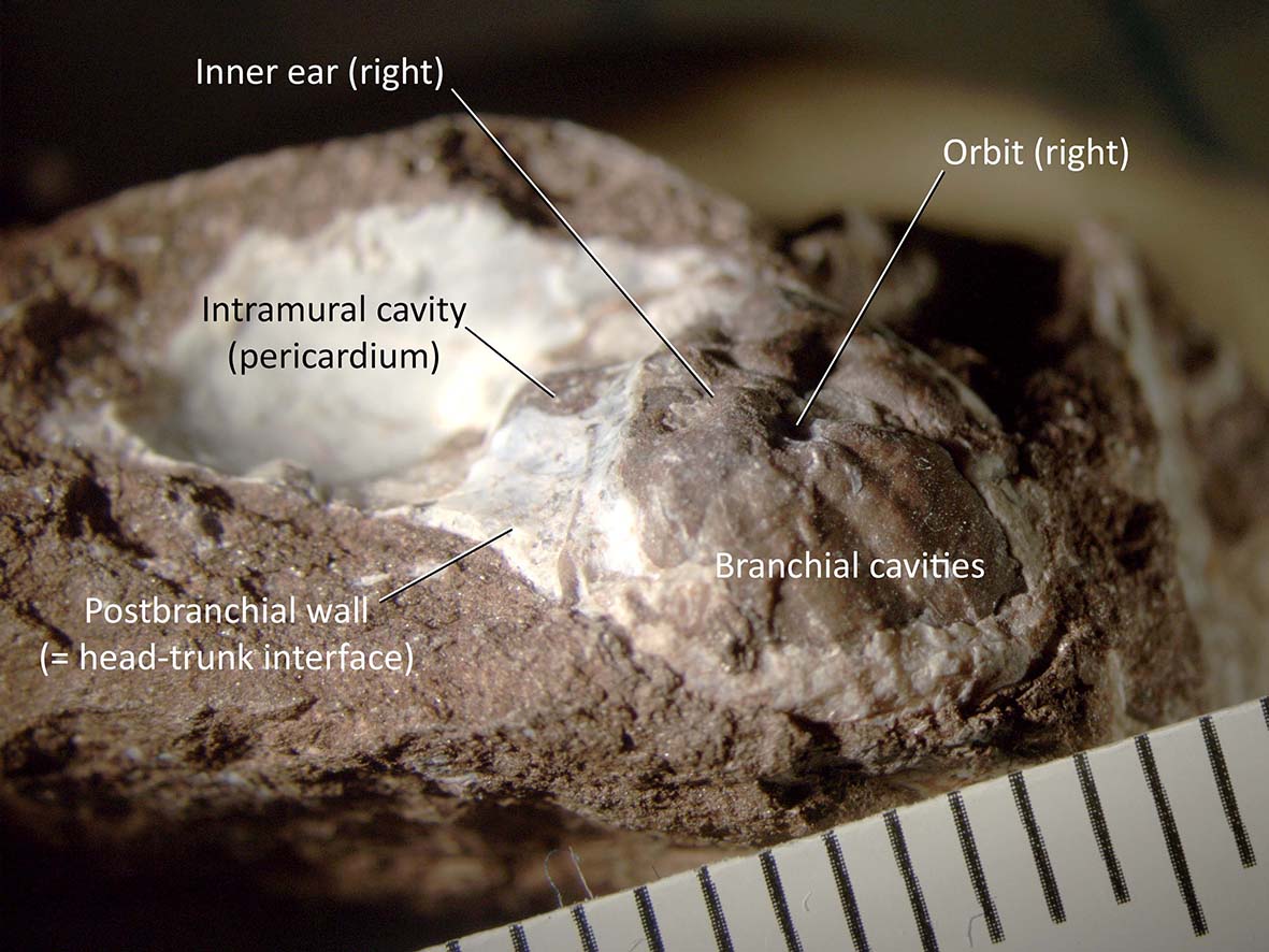
MNHN SVD 3221 (Norselaspis glacialis) in right lateral and anterior view.
- A columnar sinus superior is present (shown for the first time outside jawed vertebrates).
- The vestibular morphology implies increasingly distinct roles between the upper (semicircular canals) and lower (pars inferior) parts of the inner ear, prior to the origin of jawed vertebrates.
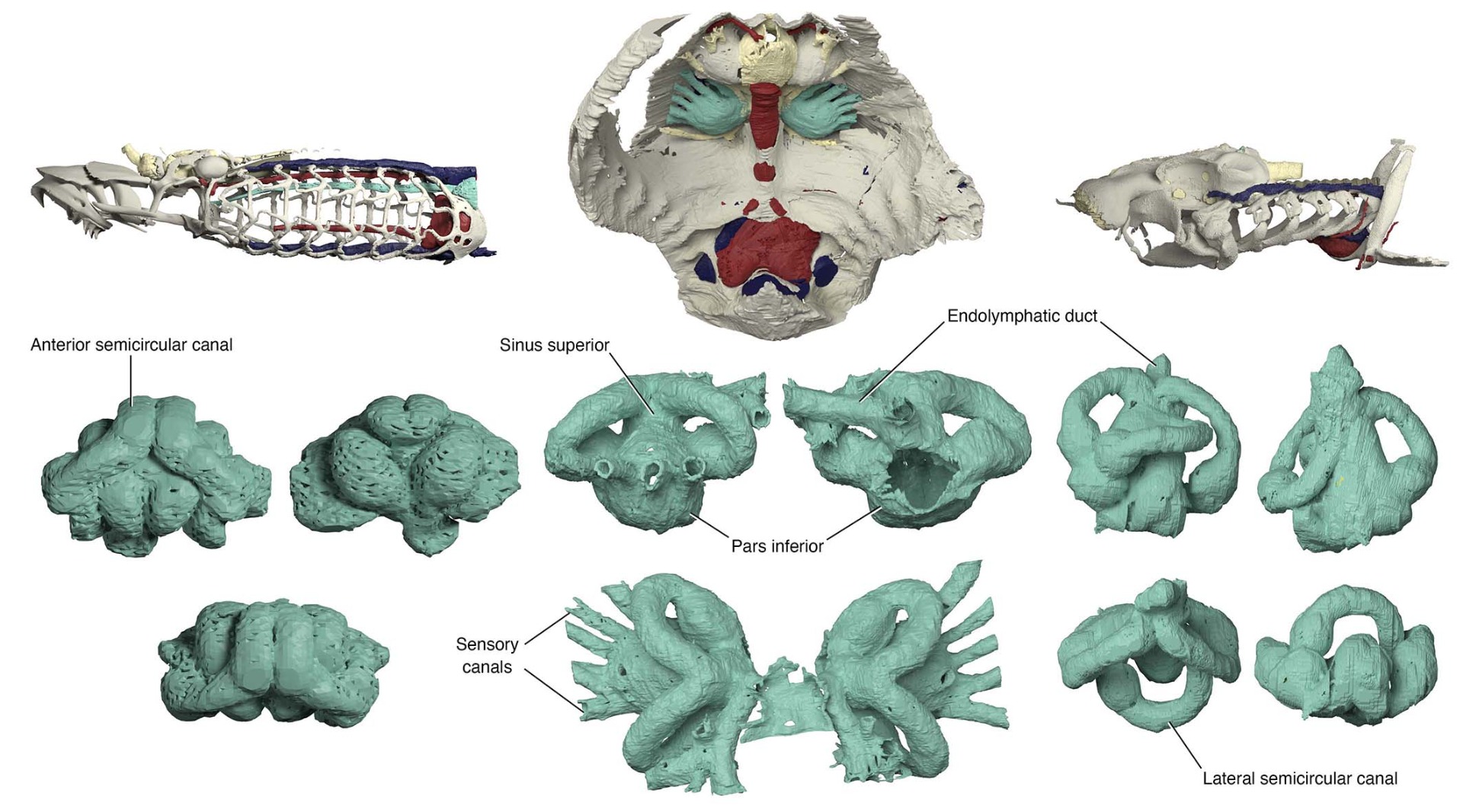
Left: The inner ear of a lamprey in (clockwise from the left top) lateral, medial, and dorsal views.
Middle: The inner ear of Norselaspis in lateral, medial, and dorsal views.
Right: The inner ear of a bamboo shark in lateral, emdial, ventral, and dorsal views, Rendering by Kristen Tietjen.
- Myodomes are preserved in the orbit, indicating that Norselaspis has seven extrinsic eye muscles.
- These seven muscles in Norselaspis correspond to those in placoderms and likely represent the ancestral configuration for vertebrates.
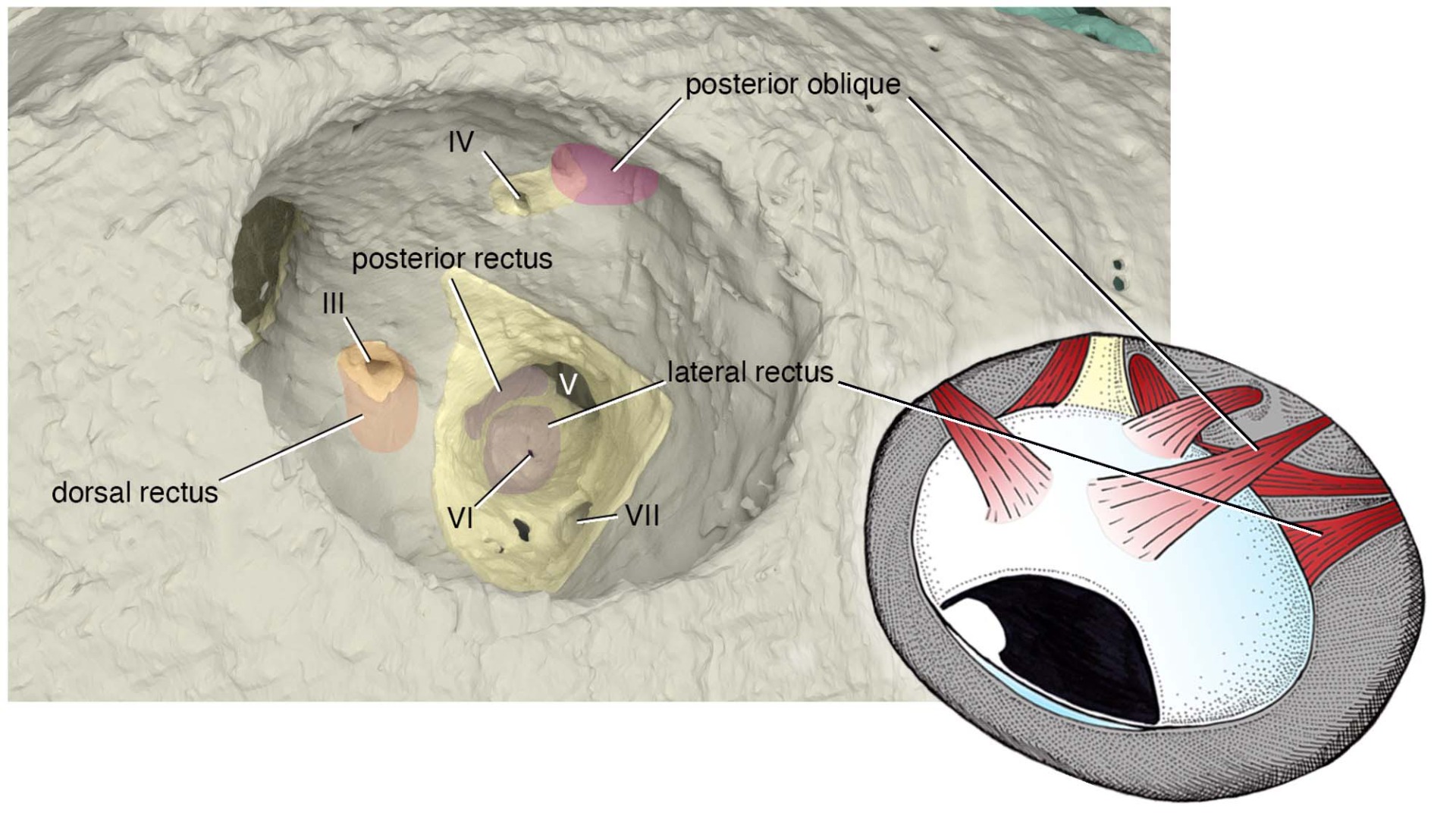
Left: The left orbit of Norselaspis with myodomes coloured by the nerves that innervate them. In anterior and lateral view. Rendering by Kristen Tietjen.
Right: Extraocular muscles of Norselaspis reconstructed in the left orbit, in dorsal view. Drawing by Tetsuto Miyashita.
- This insight resolves the unmatching homology of the six extrinsic eye muscles between lampreys and jawed vertebrates –– they each lost one muscle independently from the ancestral configuration of seven. Here fossil record shows what people considered as an anatomical constant has dynamic evolutionary history early among vertebrates!
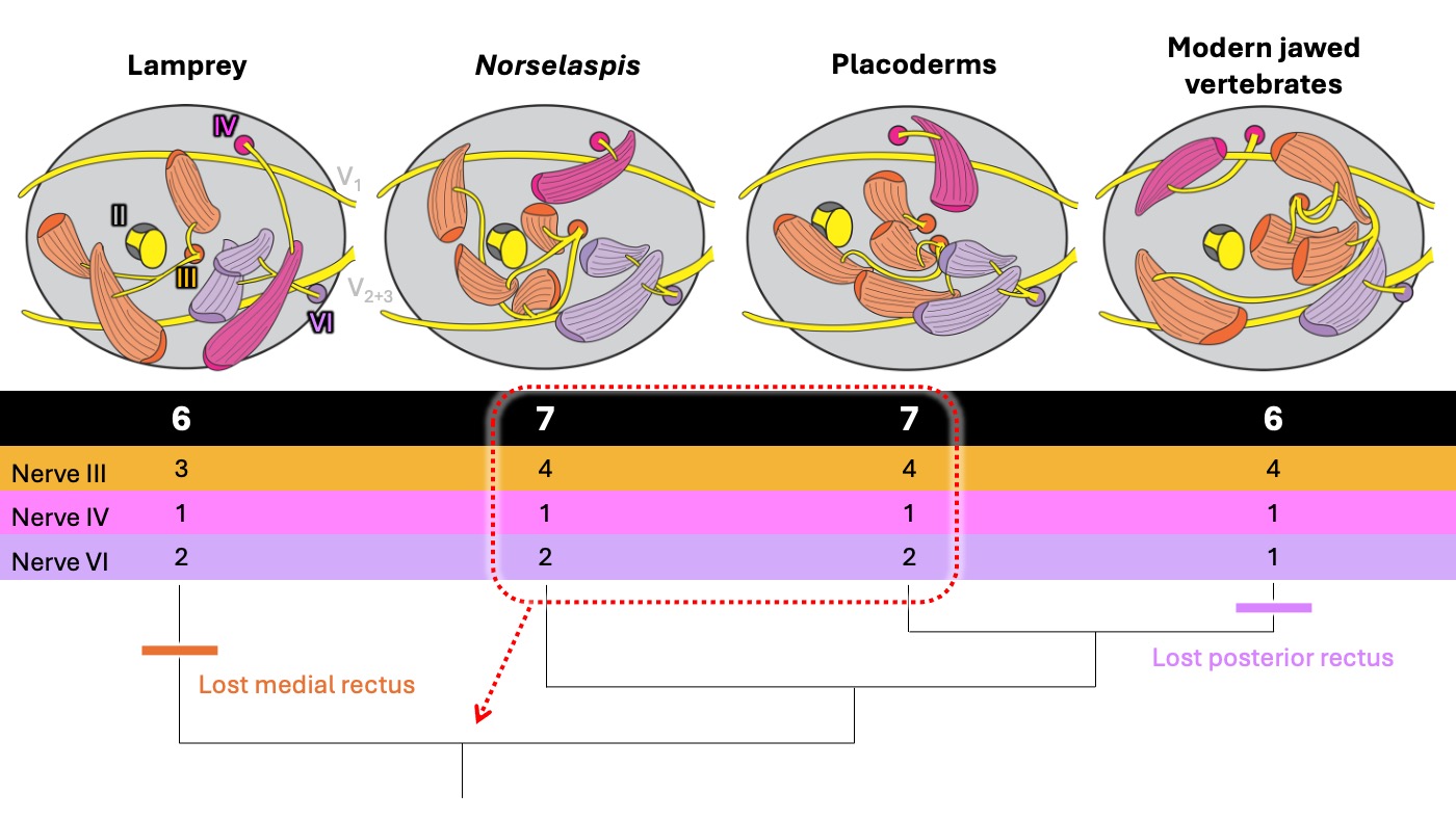
Homology and evolution of extraocular muscles across early vertebrate lineages. Diagrams show a left orbit for each lineage (anterior to the left). By innervation patterns, lampreys have a 3-1-2 pattern and modern jawed vertebrates exhibit a 4-1-1 pattern so those six muscles do not perfectly match between these lineages. Stem gnathostomes have seven muscles in a 4-1-2 pattern. With this as a primitive condition, each living vertebrate lineage lost one muscle independently.
CN III = oculomotor nerve. CN IV = trochlear nerve. CN VI = abducens nerve.
- The bony wall that separates the branchial space and the trunk in osteostracans represents the head-trunk interface that is uniquely ossified in this lineage.
- This postbranchial wall contains a pericardium.
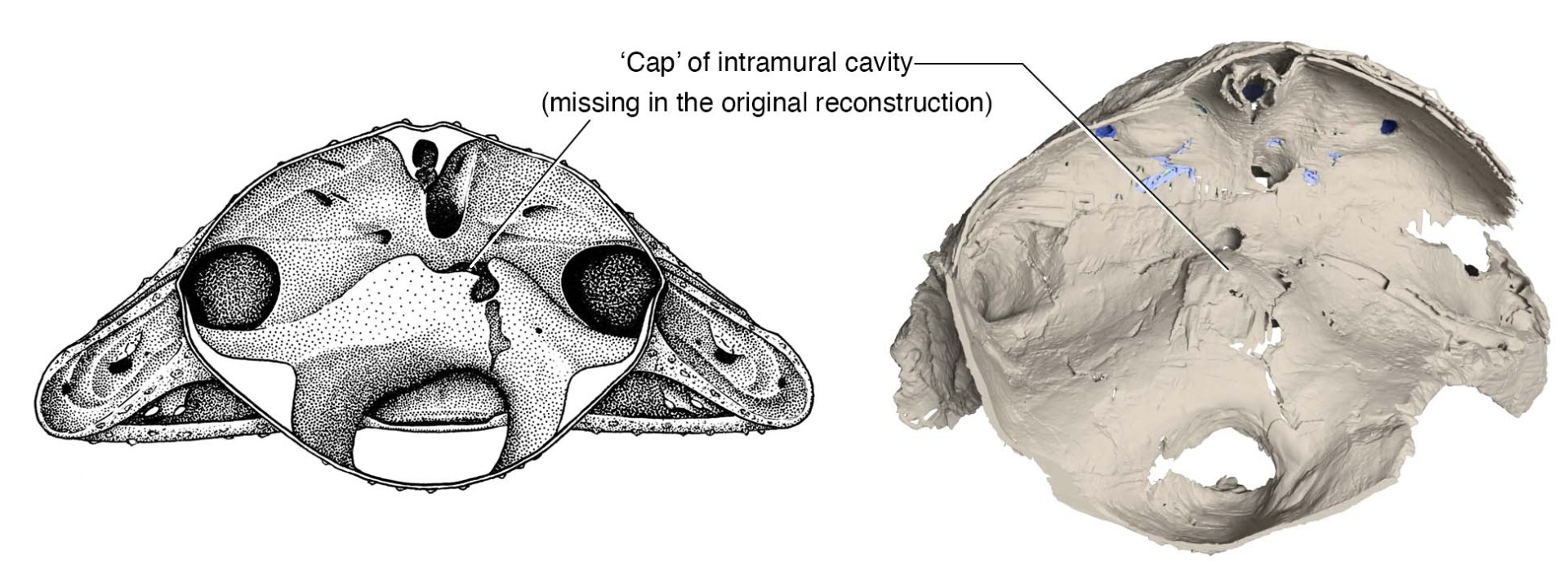
The postbranchial wall of Norselaspis in posterior view. Left: the original reconstruction (Janvier 1981). Right: new reconstruction. Note that the intramural cavity (pericardium) is closed dorsally in the new reconstruction.
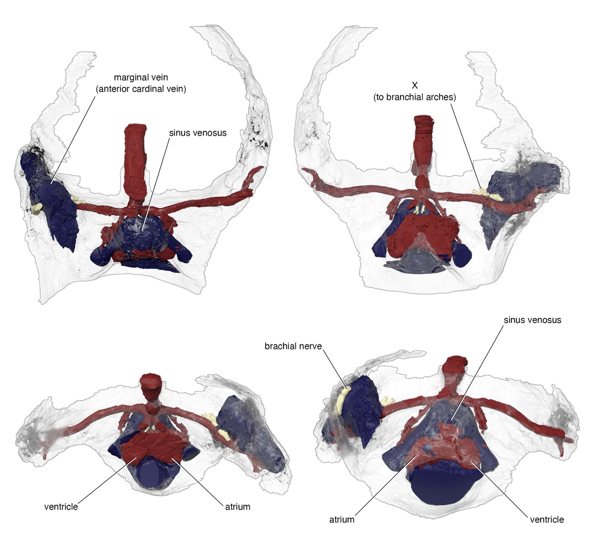
Reconstruction of the circulatory structures preserved as cavities in the bony wall that separates the head and trunk in Norselaspis, revealing the heart, major veins and arteries. Clockwise from top left: dorsal, ventral, posterior, and anterior views. Rendering by Kristen Tietjen.
- Blood drains through paired common cardinal veins like in modern jawed vertebrates. Previously, Norselaspis was interpreted to have a lamprey-like single venous drainage, but this interpretation is an artifact of damage to the original specimen.
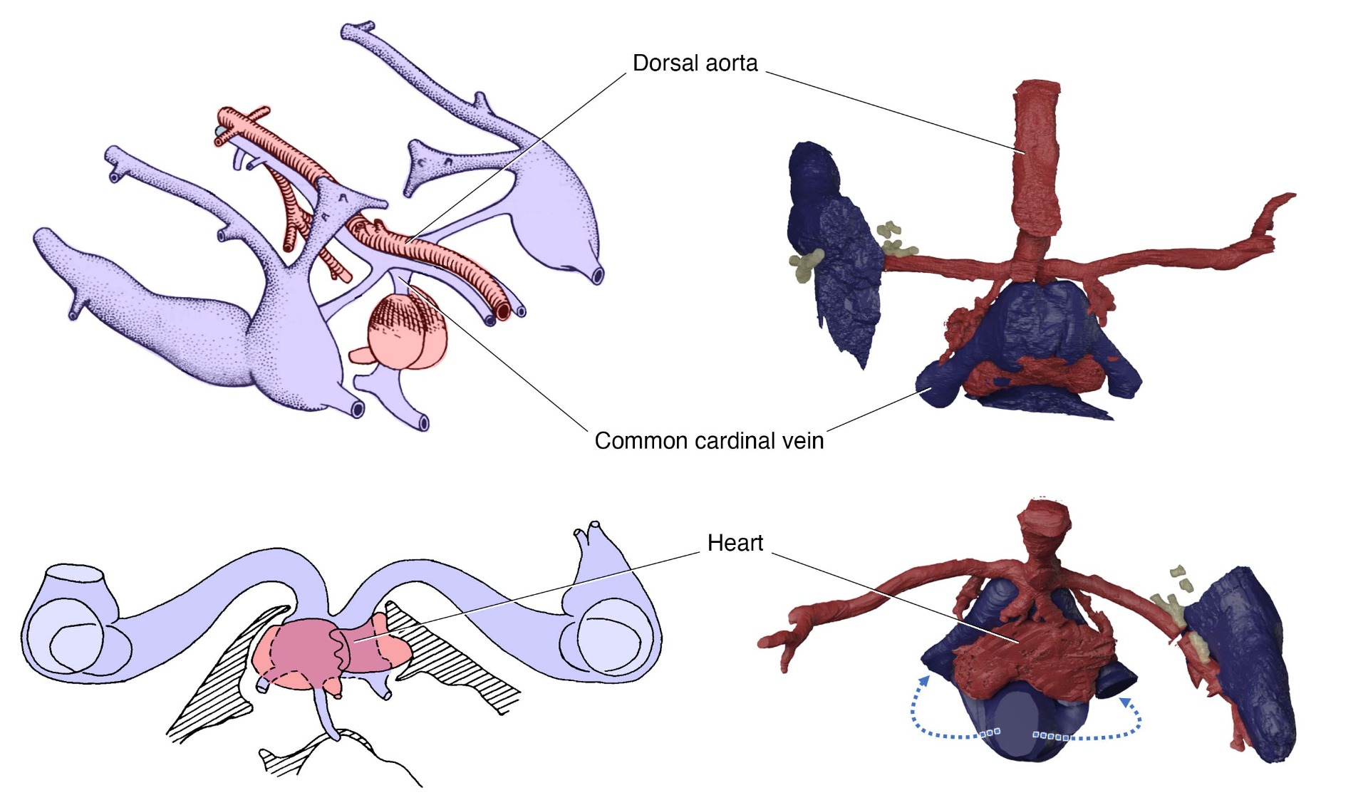
Left: the original reconstruction of the heart, dorsal aorta, and venous drainage of Norselaspis by Janvier (1981) and Janvier et al. (1991) in left oblique laterodorsal view from posterior aspect (upper) and in anterior view at the cross section through the pericardium (lower).
Right: new reconstruction of the same structures in dorsal (upper) and anterior (ower) views. Common cardinal veins are paired and each drains dorsally into the sinus venosus. Rendering by Kristen Tietjen.
- The most posterior cranial nerve (vagus nerve) innervated the pericardium via the last branchial artery (arterial pole, as in lampreys) and the extrabranchial cavity beside the common cardinal vein (venous pole, as in jawed vertebrates). The latter defines the position of the head-trunk interface.
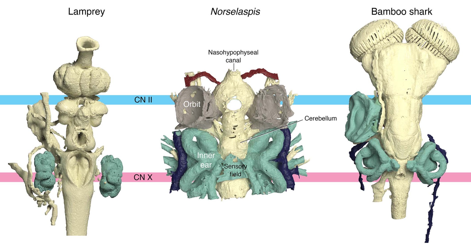
Representative vertebrate brains compared in dorsal view, aligned and scaled by spatial separation between the optic (CN II) and vagus (CN X) nerves. Rendering by Kristen Tietjen.
- The most anterior trunk nerve in Norselaspis extends to the pectoral fins. This means that Norselaspis lacks neck or throat muscles (and the nerves that innervate these structures) that in jawed vertebrates flank the head-trunk interface. *Neck muscles refer to the cucullaris, and 'throat muscles' are the hypobranchial musculature.
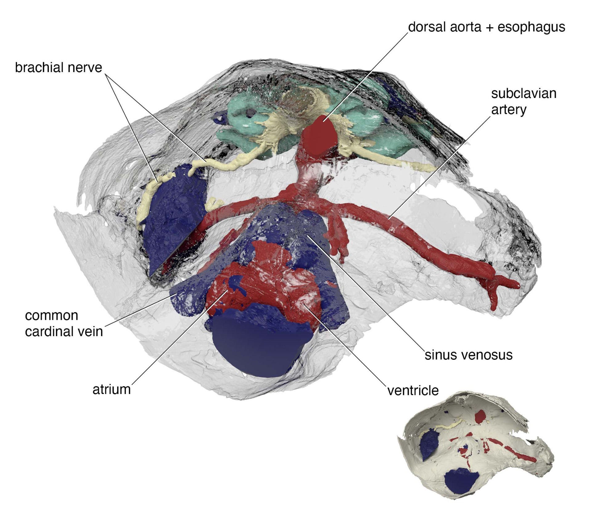
The postbranchial wall of Norselapis in oblique posterior view from right side, showing the path of the brachial nerve on the left side of the body.
- The neck and throat muscles are new structures to jawed vertebrates (no homologues among jawless vertebrates), intercalated at the head-trunk interface.
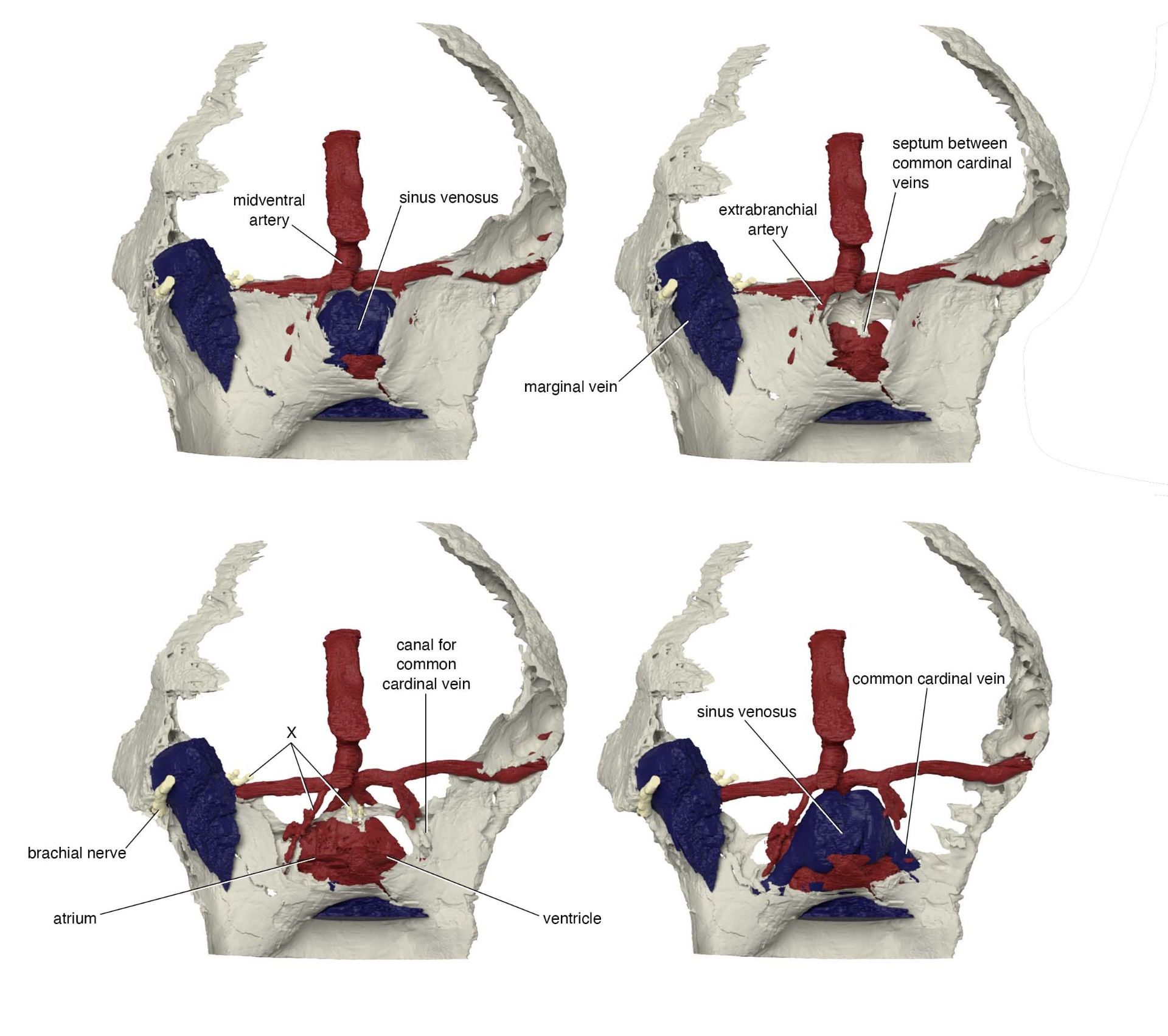
Digital dissection of the postbranchial wall of Norselapis from top to bottom, revealing the pericardium. However, there is no evidence for the neck or throat muscles. X = vagus nerve (CN X). from the left to right, it shows branchial innervation (on the medial side of the marginal sinus), cardiac branch to venous pole (paralleling the extrabranchial artery), and cardiac innervation to arterial pole (branching from the most posterior [eighth] branchial projection, paralleling the eighth efferent branchial artery). Rendering by Kristen Tietjen.
- This also implies: the muscle precursors that contribute to these muscles in jawed vertebrates instead formed part of the pectoral fin. These muscles perhaps arose as serial homologues.
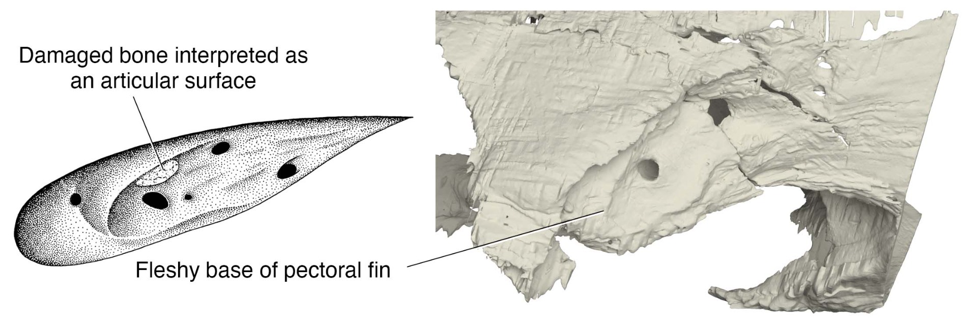
Left: The original reconstruction of the pectoral fenestra in Norselaspis posited an articular cartilage for an endoskeletal joint with pectoral fin. From Janvier (1981).
Right: New reconstruction shows that this was a damage to the perichondral bone. Norselaspis had a fleshy base for its pectoral fin. Rendering by Kristen Tietjen.
- A pectoral fin attachment in osteostracans was posited to have an endoskeletal joint. We showed in Norselaspis this is an artifact of damage to the original specimen. There is no evidence for any endoskeletal joints in ostracoderms. The pectoral fin had a fleshy base.
- The implication is that joints evolved with the origin of vertebrate jaws.

Life reconstruction of Norselaspis by Kristen Tietjen.
- Pectoral fin attachment is tilted so abductors occupy greater surface area than adductors. The angle and configuration of the finbase implies function in increasing drag for improved maneuverability, including thrust and lift.
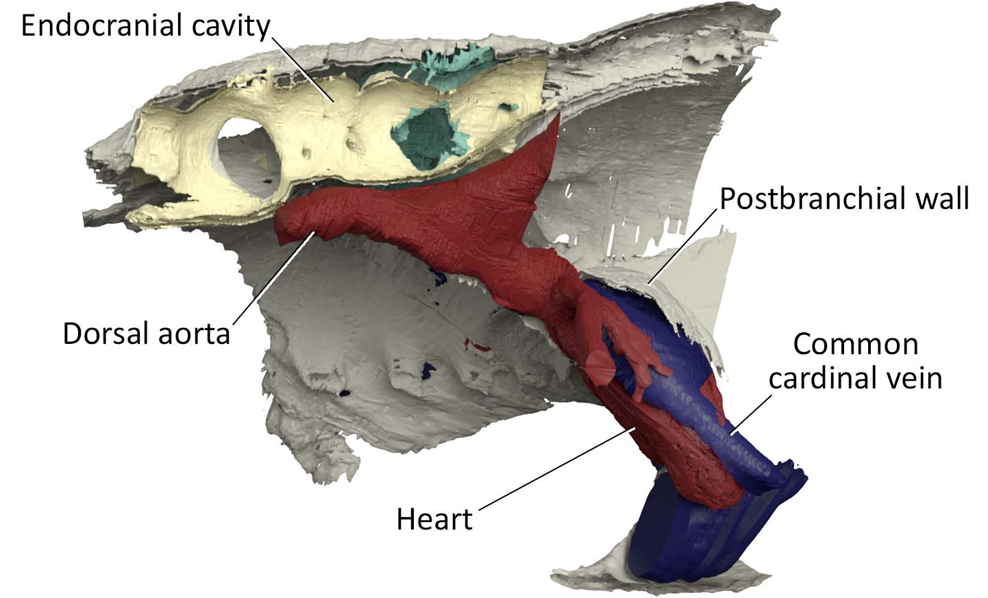
Sagittal 'cut-away' view of the postbranchial wall and endocranial cavity of Norselaspis. The postbranchial wall corresponds in jawed vertebrates to the neck and hypobranchial muscles. Rendering by Kristen Tietjen.
- The head-trunk interface did not shift in position from jawless to jawed vertebrates., but new structures (neck and hypobranchial muscles) were intercalated, flanking the interface.
- This means that the shoulder girdle (or pectoral appendages) do not have their evolutionary origin in the pharyngeal domain.
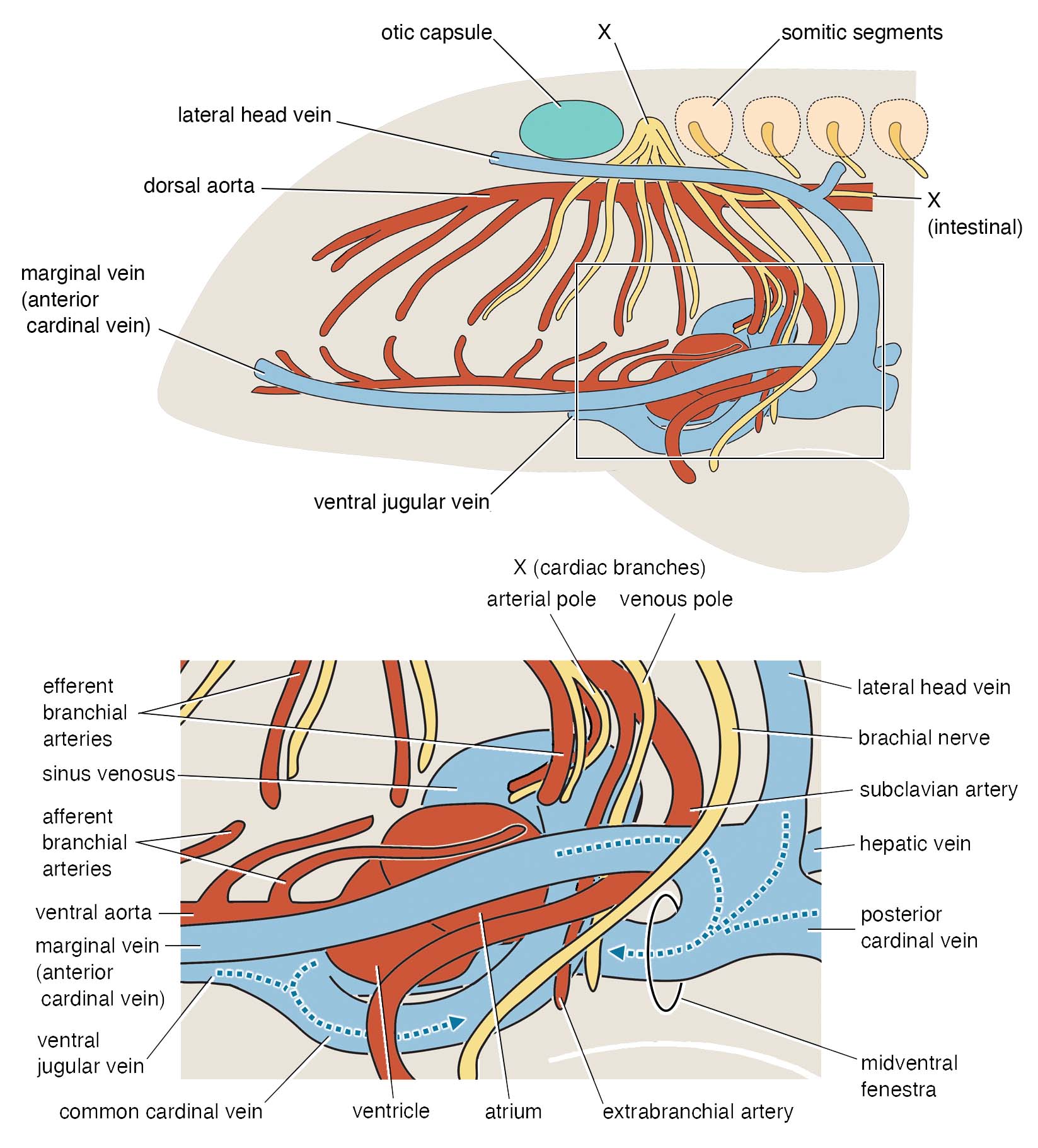
Schematic drawing for the anatomy of the head-trunk interface in Norselaspis. Remarkably, all anatomical structures depicted here have osteological correlates identified in MNHN SVD 3221, even down to the epaxial-hypaxial differentiation implied for the innervation of pectoral muscles by the brachial nerve. Illustration by Tetsuto Miyashita.
- Both evolutionary and developmental histories are complicated for structures on either side of the head-trunk interface. Multiple cell lineages migrate and interact across the head-trunk interface (e.g., second heart field, precursors for hypobranchial muscles, vagal neural crest cells). This complex spatial relationship can be further complicated as different vertebrates have different relative dimensions of the flanking domains. In osteostracans, positions of the arteries, nerves, and muscles are shifted laterally by the expansion of the branchial cavities.
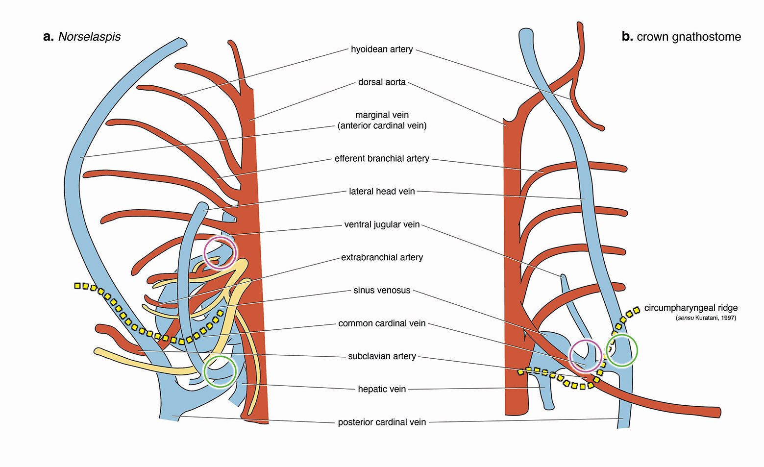
Selected nerves and vasculature of Norselaspis (left) and a modern jawed vertebrate (right) viewed from the top. The yellow broken line shows the boundary between the head and trunk. Pectoral fins form behind this line (=in the trunk domain) in both lineages.
Circles indicate morphologically comparable points of venous drainage between the two forms.
For Norselaspis, the most posterior cranial nerve (head domain) and the most anterior spinal nerve (trunk domain) are shown. The former sends its branches to the heart and intestine.
Illustration by Tetsuto Miyashita.
- But the original spatial coordinates are conserved along the body axis. Based on this information, osteostracans already had a gnathostome-like assembly of a head-trunk interface, except for lacking the neck and hypobranchial muscles.
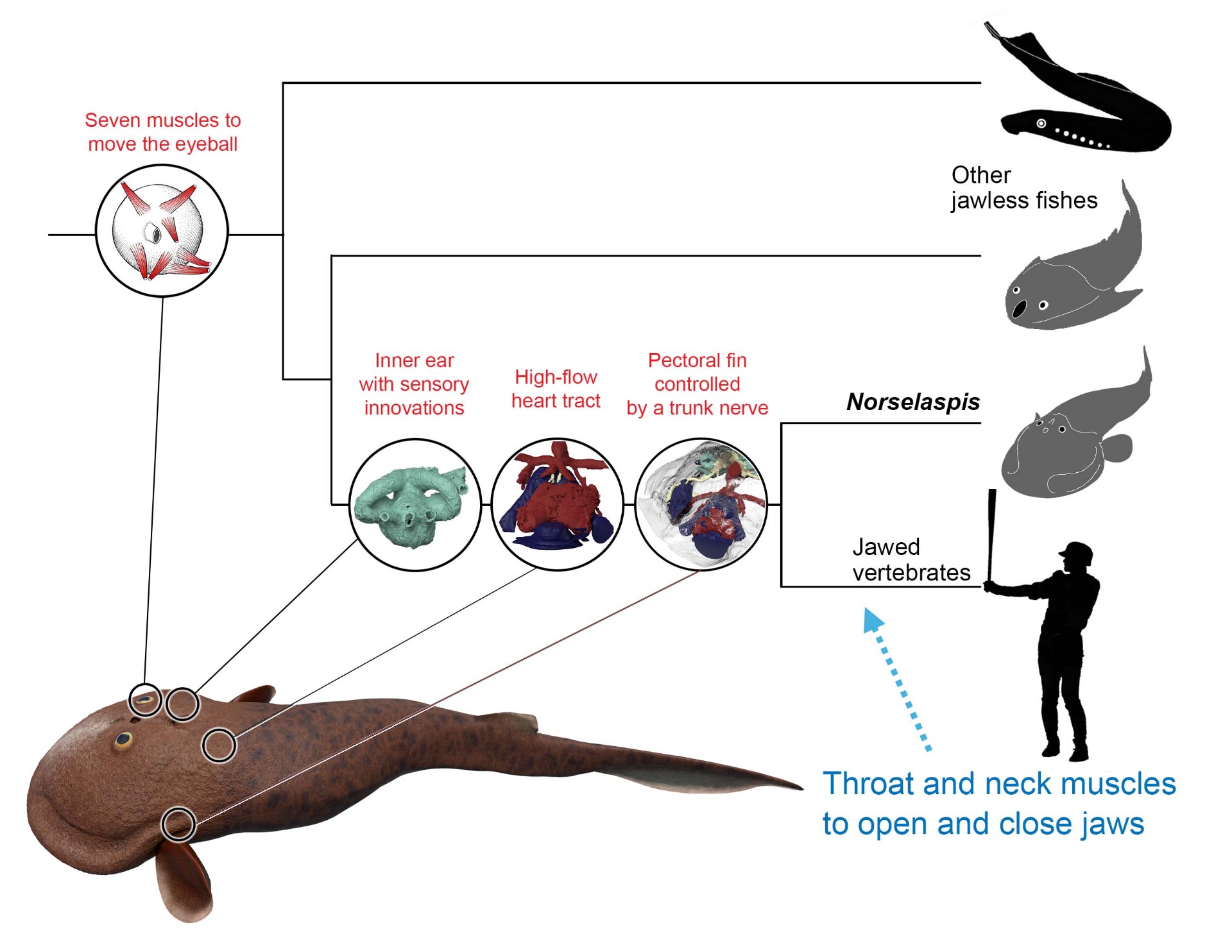
Many derived traits of jawed vertebrates have deep origins among common ancestors between Norselasps and us. Elaborate sensory systems, increased cardiac output, and greater locomotory control have been attributed as innovation by the earliest jawed vertebrates, but our work shows that they preceded jawed vertebrates. What is truly novel for jawed vertebrates includes jaws (of course) and the muscles in the neck and throat that help opening and closing of the mouth, now possible because the shoulder girdle is decoupled from the skull.
- With sensory innovations identified in the eyes and inner ears oNorselaspis, increased cardiac output implied from its heart size and venous configuration, and greater locomotory control inferred from the pectoral anatomy, many derived gnathostome traits have a deep root in the jawless stem segment, among common ancestors between osteostracans and jawed vertebrates.
- Thus, jaws are a follower to these character changes than a facilitator/functional driver for them. This improved understanding of preconditions to the origin of jaws generate two important insights.
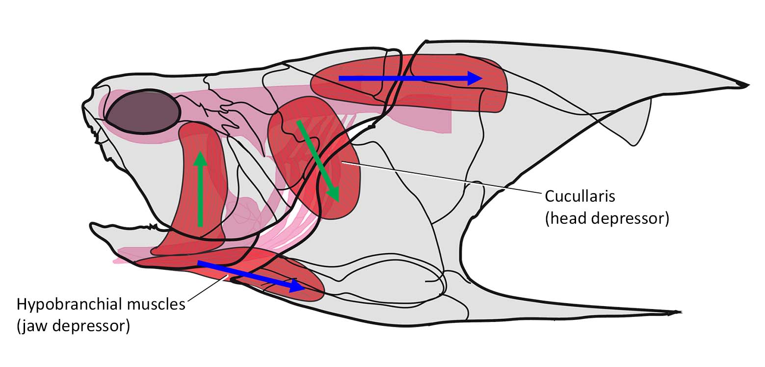
An arthrodire placoderm (Coccosteus cuspidatus) with major head and trunk muscles reconstructed. Cucularis and hypobranchial muscles each function in depressing the head (helps closing the mouth) and depressing the jaw (helps opening the mouth) respectively. Original illustration by Miles and Westoll (1968), modified by Miyashita (2016).
- First, these derived traits in Norselaspis shorten the branch length (reduce the number of character changes optimized) between osteostracans and jawed vertebrates. The morphological gap between jawless and jawed vertebrates is narrower than conventionally predicted.
- Second, the standard scenario for the origin of jaws as a linear progression to macropredators is flawed. Fish got 'faster and smarter' well before they acquired jaws. We consider an alternative scenario: the earliest jaw skeletons evolved to extend an active lifestyle already supported by sensory, circulatory, and locomotory innovations that we identified in Norselaspis.
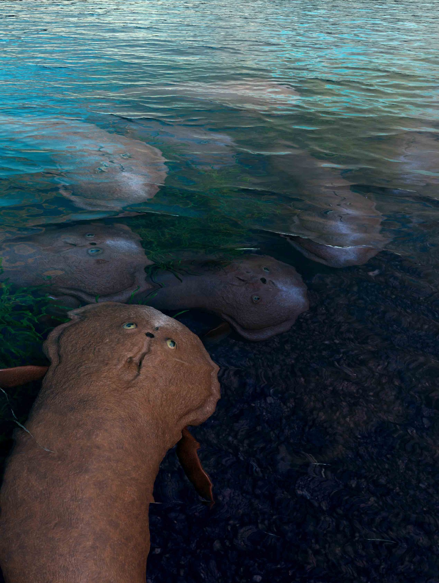
HEADS UP: A school of Norselaspis glacialis rise to the surface in what is now the Arctic Archipelago of Svalbard, Norway, 412 million years ago. Illustration by Kristen Tietjen.
- Under this scenario, the jaw (with its hinge joint) and the muscular neck and throat arose in tandem and facilitated a substantial increase in feeding current generated by the mouth. Only then, the greater effciencies in suction feeding drove ecological diversification that characterizes the early radiation of jawed vertebrates.
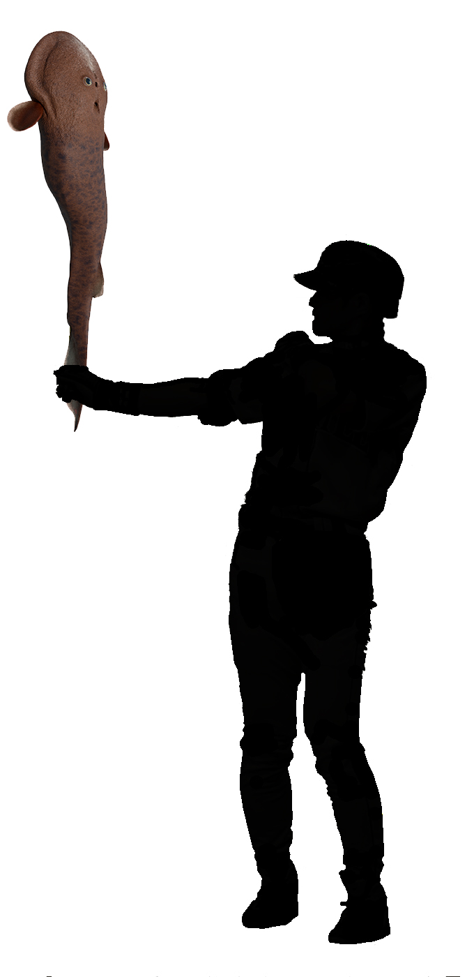
Although an osteostracan cannot play baseball, maybe it is possible to play with it. This is the ultimate torpedo bat, which may not have been suitable for a contact hitter like Ichiro Suzuki.
Life restoration of Norselaspis by Kristen Tietjen. Drawing by Tetsuto Miyashita.
